Images from:
(1983d) King, D.G. and M.A. Tanouye Anatomy of motor axons to direct flight muscles in Drosophila. Journal of Experimental Biology 105: 231-239. [email for PDF: dgking@siu.edu]
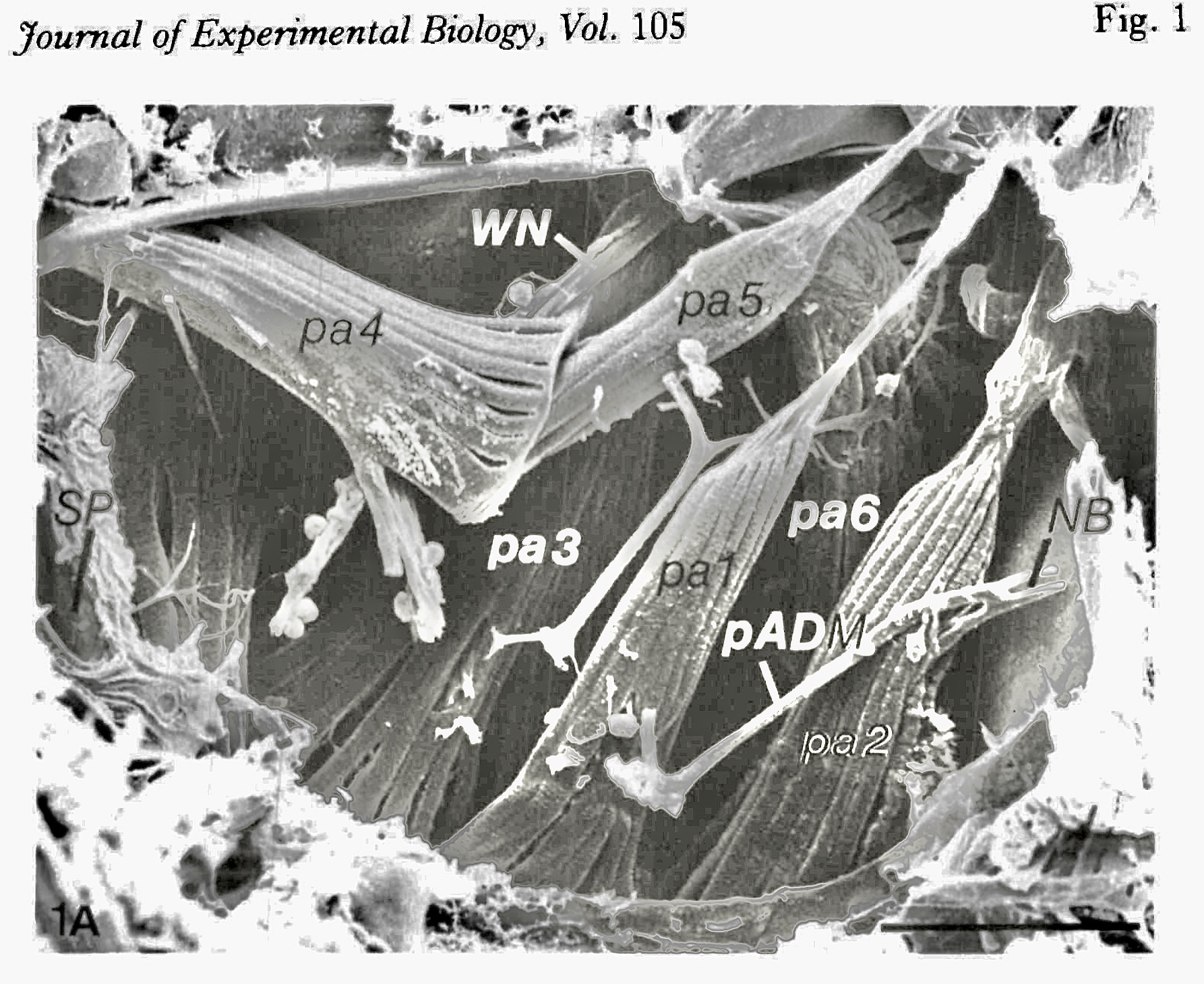
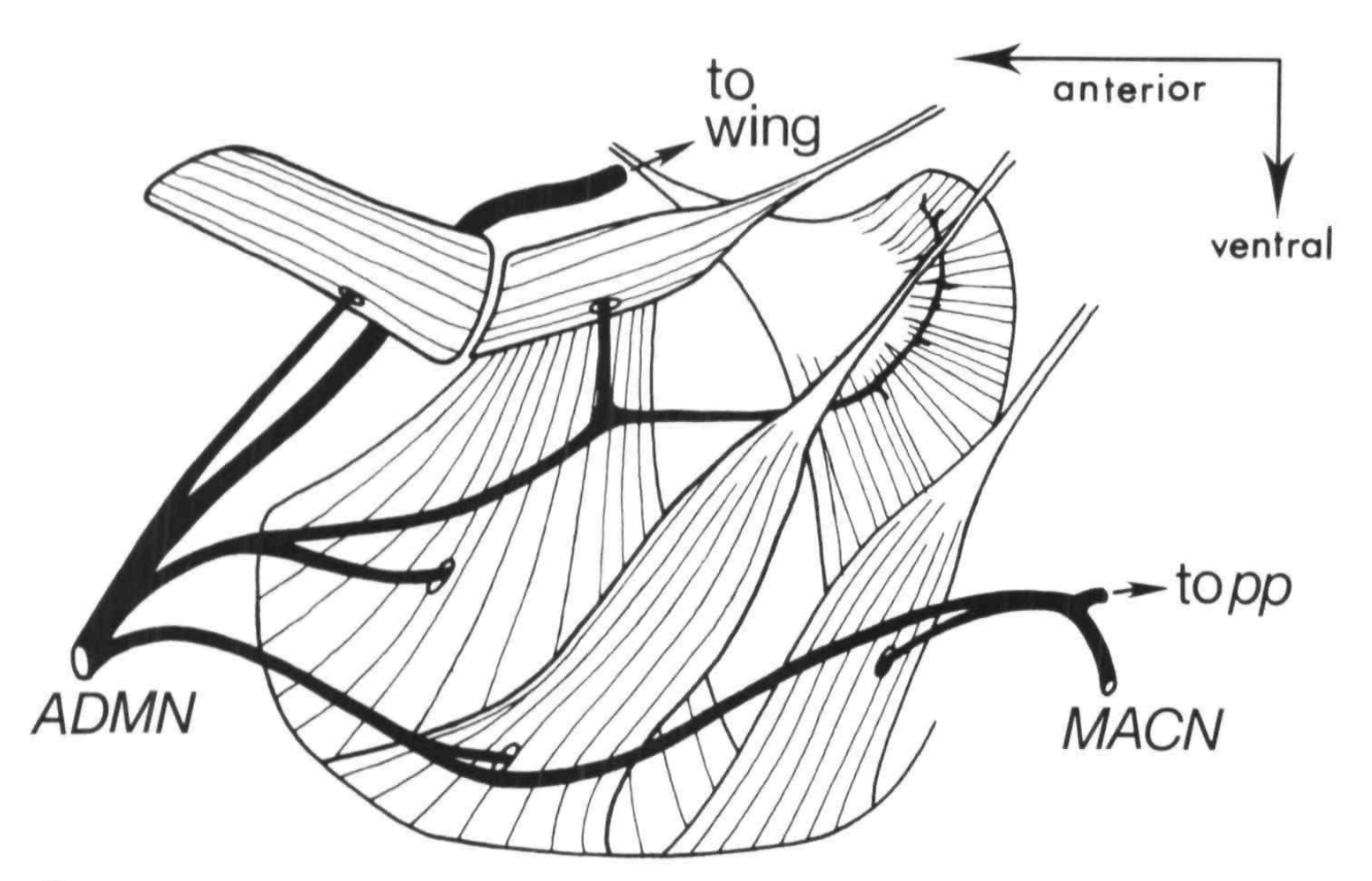
SEM image of Drosophila direct flight muscles, viewed from inside the right thoracic wall (anterior to left). Structures labelled pa are anterior pleural muscles; WN = wing nerve; pADM = posterior branch of anterior dorsal mesothoracic nerve; NB = nervenbrucke connecting pADM with the mesothoracic accessory nerve; SP = spiracle. Diagrammatic representation of direct flight muscles. ADMN = anterior dorsal mesothoracic nerve; MAC = mesothoracic accessory nerve; pp = posterior pleural muscles. Parasagittal section of Drosophila thorax (anterior to left). Arrowheads indicate locations for electron micrographs in Fig. 2 B, C, D, E; Structures labelled pa are motor axons to anterior pleural muscles; DVM = dorsoventral direct flight muscle; TTM = tergotrochanteral muscle; WN = wing nerve; ADMN = anterior dorsal mesothoracic nerve; HN = haltere nerve.

Comments and questions: dgking@siu.edu
SIUC / Zoology / David King
https://dgkinglab.siu.edu/King&Tanouye-images.htm
Last updated: 21 October 2023 / dgk